Introducing TrueLearn
Updated: 03/17/2016
At the PAEA Summit I recently attended in Washington D.C. I learned that PA students and practicing PAs should understand that even once we graduate, we will always be a student. Continuous learning is an expectation not just of our peers or NCCPA, but of our employers as well, and our colleagues (physicians and NPs). Collectively, we must work to ensure we are practicing at the top of our career, implementing the best practices and presenting the most up-to-date information to our patients. One colleague put it plainly, “either be at the table, or be on it.” This can be interpreted in many ways, but I believe that regular review of content keeps us competitive.
Over the next 3 months, I will be selecting topics that are most commonly tested by the NCCPA and PAEA for exam preparation. TrueLearn, which provides next generation question banks (called SmartBanks), will provide free practice questions on these topics to provide insight into their test preparation expertise. One of the best ways to prepare for your exams is through quality question banks. TrueLearn has introduced an incredible opportunity to work with the PA-student and PA community to help us excel in our didactic education and clinicals, and on our boards. Moving forward, I am pleased to announce TrueLearn to the table of exam preparation for PAs from the start of your education. See below for our first post discussing Heart Murmurs. This topic is important as Cardiology makes up 16% of the PANCE and PANRE exams.
SmartBanks, brought to you by TrueLearn, the nation’s most trusted test preparation, provide the highest level of test readiness available today.Their products have served up over 39 million practice questions in 2015 alone, achieving an 11% score improvement after using their SmartBanks. TrueLearn’s SmartBanks have been utilized for USMLE, COMLEX, and specialty board exams in general surgery, anesthesiology, emergency medicine, family medicine, pediatrics, OB/GYN, internal medicine, and psychiatry. These exams are written by the nation's leading specialists and are used by most medical students and residents in preparation for shelf (clinicals) and board exams.
TrueLearn’s SmartBanks include specialized medical content crafted to mirror rotation exams, the PACKRAT, the PANCE and the PANRE. Why stop there? Use these exams to help you prepare for Certificates of Added Qualification (CAQ) - the limits are endless!
Visit TrueLearn.com/Physician-Assistant/ today to learn more about their physician assistant products.
TrueLearn’s SmartBanks provides:
- An accurate reflection of what you can expect on test day, both in regard to content and difficulty, so you can be rest assured you are prepared
- A realistic test interface that closely mimics features provided for your rotation exams, PANCE, or PANRE. Alas, a testing platform that you can utilize from PA student on clinical rotations to clinician.
- Board-style vignettes reflective of the rotation exams, PANCE and PANRE
- Explanations with updated references, learning points
- User-friendly software with easily accessible IT support for questions or concerns with quick 24-48 hour response
- Adaptive performance analytics with personalized learning - fit to meet your weaknesses and allowing you to see where your strengths exist so you can focus your learning
- Comparative analysis to your nationwide peers on individually created exams and questions
- Train more effectively and monitor your progress as you go with a chart of your performance
- No more purchasing question banks with questionable validity - questions are written by PAs and MD/DOs and tested for accuracy, ensuring the level of difficulty will prepare you for test day
- Prepare, assess, and improve your performance from home or on the go using the mobile app available on all smart devices
Disclosure: I am not an employee of TrueLearn.
Heart Murmurs
Key features
|
Description
|
Radiation
|
Other Clues
| |
Mitral Valve Prolapse: Regurgitant flow across mitral valve
|
Decreased left ventricular volume results in earlier prolapse and click heard earlier in systole, closer to S1
Increased left ventricular volume results in delayed prolapse, click heard later in systole
|
Mid-systolic click followed by a mid-to-late systolic murmur and loud S2
“click” is caused by prolapse of leaflets into left atrium and tensing of mitral valve apparatus
|
Maneuvers that increase left ventricular end diastolic volume (preload): squatting from seated or standing position
Maneuvers causing delay in prolapse: handgrip, standing from seated position, valsalva
|
Leads to mitral regurgitation
Predisposed to infective endocarditis
ADA no longer recommends prophylactic antibiotics for MVP
|
Mid-systolic (HAPI) Murmurs
Key features
|
Description
|
Radiation
|
Other Clues
| |
Hypertrophic Cardiomyopathy
|
Will cover this topic at a future date
| |||
Aortic Stenosis
|
Paradoxical split S2
Elderly, syncopal episodes
|
Crescendo-decrescendo harsh systolic ejection click radiating to carotids
Best heard on right
Brisk upward deflection of carotid
|
Base of heart
Severe: can be heard near carotid arteries
|
EKG: Left ventricular hypertrophy
|
Pulmonary Stenosis
|
Congenital disorder
Left sided HF: DOE, jugular pulsation, parasternal lift
|
Preceding systolic sound during expiration
Systolic murmur heard best at 2nd-3rd LICS, radiates to left shoulder
|
EKG: right axis deviation
| |
Innocent Murmur
|
Will cover this topic at a future date
| |||
Holosystolic (MTV) Murmurs
Key features
|
Description
|
Radiation
|
Other Clues
| |
Mitral Regurgitation
|
Chronic mitral regurgitation seen in cases of acute rheumatic fever, but presents in first-second decade
|
High-pitched, blowing
Holosystolic murmur radiating to left sternal border
|
Apex (best) → Axilla
Reduced with valsalva
|
EKG: left axis deviation or LVH
|
Tricuspid Regurgitation
|
S3 may be present
|
Holosystolic murmur heard best at right sternal border, 5th ICS
NO EJECTION CLICK
Opening snap + diastolic murmur
|
Left sternal border
Increased with inspiration
|
EKG: right axis deviation
|
Ventricular Septal Defect (VSD)
|
Most common of all congenital heart defects
Depends on size of defect - from asymptomatic to CHF
|
Systolic murmur at LLSB
|
Diastolic (ARMS) Murmurs
Key features
|
Description
|
Radiation
|
Other Clues
| |
Aortic Regurgitation
|
Younger pt with h/o Marfan’s syndrome
or older patient
|
High-pitched Blowing or rumbling diastolic decrescendo-crescendo (“V”) murmur heard at left sternal border or apex of heart
Bounding “water hammer” pulse
|
Classic: diastolic murmur at RSB due to backflow of blood across aortic valve
|
Rumbling sound at apex (Austin Flint Murmur) - retrograde blood across aorta mixes with anterograde blood from left atrium
|
Mitral Stenosis
|
Enlarged left ventricle
Common after rheumatic fever
Hemoptysis, presents in 30-40s
|
(1) Loud first heart sound: wide closing excursion of leaflets
(2) Prominent P2 of second heart sound (split S2): elevated pulm-art pressures
(3) Opening Snap: sudden tensing of leaflets after they’ve completed their opening excursion
(4) Mid-diastolic rumble - increased flow across stenotic MV during atrial contraction
|
Opening snap followed by low pitched diastolic rumble
|
Leads to pulmonary hypertension and heart failure → high LA pressures = high pulmonary vasculature and right heart pressures
|
Valvular Heart Disease
- Aortic Stenosis - narrows valve opening, impeding the ejection function of the left side of the heart (most common valvular disease in US)
- Thready carotid pulse
- Aortic Regurgitation (insufficiency) - volume overloading due to retrograde blood flow into the left ventricle
- Bounding pulses and wide pulse pressures
- Mitral Stenosis - impedes blood flow between left atrium and ventricle
- Mitral Regurgitation (insufficiency) - retrograde blood flow and volume overload of the left atrium
- Mitral valve prolapse - may be asymptomatic, but associated with MR; mostly thin females with minor chest wall deformities, midsystolic clicks, and late systolic murmur
- Most common causes of mitral/aortic valve disorders are congenital defects; other causes include rheumatic heart disease, connective tissue disorders, infection, sensile conditions
- Most common presenting symptoms: dyspnea, fatigue, decreased exercise tolerance
- Other: Cough, rales, paroxysmal nocturnal dyspnea or hemoptysis, hoarseness
- Physical exam - heart murmur +/- palpable thrill
- Diagnostic Studies
- EKG - not useful for specific diagnosis → shows chamber hypertrophy
- CXR
- Aortic → left sided atrial enlargement, ventricular hypertrophy
- Mitral → atrial enlargement only
- Echo (transesophageal) and cardiac cath → definitive
- Treatment
- Surgical repair or replacement of defective valve
- Good exercise tolerance → diuretics and vasodilators for pulmonary congestion and digoxin or BB for dysrhythmias
- Anticoagulant therapy for thromboemboli prophylaxis
- Antibiotics to prevent endocarditis and recurrent rheumatic fever
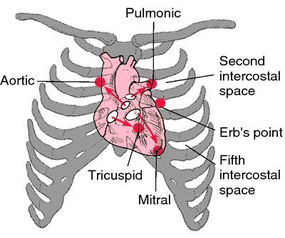
Location
|
Radiation
|
Intensity
|
Pitch/Quality
|
Aids to Hearing
|
Associated findings
|
Timing
| |
Aortic Stenosis
|
2nd Right-ICS
|
Neck and LSB
|
Loud with a thrill
(Grade 4-6)
|
Medium/
Harsh
|
Patient sitting and leaning forward
|
Midsystolic
| |
Aortic Regurgitation
|
2nd-4th Left-ICS
|
Apex and RSB
|
Grade 1-3
|
High pitch/
Blowing
|
Patient sitting and leaning forward
Full exhalation
|
Mid-systolic or Austin Flint murmur suggests large flow;
arterial pulses large and bounding
|
Systolic (soft) and decrescendo in early diastole
|
Mitral Stenosis
|
Apex
|
Little/None
|
Grades 1-4
|
Low pitch
|
Patient in LLD
Full exhalation
|
S1 accentuated;
opening snap follows S2
|
Mid-Diastolic
|
Mitral Regurgitation
|
Apex
|
Left axilla
|
Soft to loud
|
Medium to high pitch;
Blowing
|
N/A
|
S2 decreased;
apical impulse prolonged
|
Pansystolic
|
Tricuspid Regurgitation
|
LLSB; holosystolic
|
Right sternum and xiphoid area
|
Variable
|
Medium;
Blowing
|
Increases with inspiration
|
JVP elevated
|
Pansystolic
|
Pulmonic Stenosis
|
2nd-3rd Left-ICS;
Mid-systolic
Crescendo
-Decrescendo
|
Left shoulder and neck
|
Soft to loud, possibly associated with thrill
|
Medium;
Harsh
|
N/A
|
Early pulmonic ejection sound (common)
|
Mid-Systolic
|
Mitral Valve Prolapse
|
Late systolic murmur
|
Mid-systolic
|
Tricuspid and Pulmonic Valve Disorders
- Present during early infancy or childhood; adults present with stenosis from rheumatic scarring or connective tissue disease
- Tricuspid regurgitation is intrinsic or functional
- In all cases, right sided pressure overload → right sided cardiomegaly, systemic venous congestion, and right sided heart failure
- Clinical features: exercise intolerance
- JVD, peripheral edema, hepatomegaly
- Diagnostic Studies
- CXR: prominent R-heart border, dilation of SVC
- EKG: right axis deviation, p-wave abnormalities with R-atrial enlargement, prominent R and deep S waves of RVH
- Treatment
- Sodium restriction, diuretic therapy
- Surgical repair, valvuloplasty or replacement
Cyanotic Defects
Tetralogy of Fallot
|
6-10%
|
Crescendo-decrescendo holosystolic at LSB, radiates to back
1. Right ventricular hypertrophy
2. Pulmonic Stenosis
3. Overriding aorta
4. VSD
Tet-spells: irritability, tachypnea, cyanosis worse with exertion (feeding or intense crying)
Reduced with squatting: increases SVR
|
R → L shunting
Cyanosis, blubbing, increased RV impulse at LLSB,
Loud S2
|
Polycythemia; tet spells (hypercyanotic) include extreme cyanosis, hyperpnea, and agitation
EMERGENCY
|
Pulmonary Atresia
|
1-3%
|
Depends on presence of tricuspid regurgitation
|
Cyanosis with tachypnea at birth, tachypnea without dyspnea, hyperdynamic apical pulse, Single S1/S2
|
Sudden onset severe cyanosis and acidosis
EMERGENCY
Indomethacin
|
Hypoplastic Left Heart Syndrome
|
7-9%
|
Variable; not diagnostic
|
Shock, early heart failure, respiratory distress
Single S2
|
More often in males, 25% of cardiac death before age 7
|
Transposition of the great Vessels
|
5-7%
|
Systolic murmur if associated with VSD; systolic ejection murmur if associated with pulmonary stenosis
No exchange between right and left circulation
|
Cyanosis in newborn (MC)
Tachypnea without respiratory distress
If large, sx of CHF and poor feeding
Single loud S2
Absent LE pulses if aortic arch obst
|
Prostaglandin!!
|
Non-Cyanotic Defects: All Ventricles Provide Circulation (AVPC)
Atrial Septal Defect
|
7% of all defects
Cause: patent foramen ovale
|
Systolic ejection murmur at 2nd LCIS
Early to mid systolic rumble
|
Failure to thrive, fatigue
RV heave, wide fixed split S2
| |
VSD
|
Most common of all congenital heart defects
|
Systolic murmur at LLSB
|
Depends on size of defect - from asymptomatic to CHF
|
Outlet VSDs more common in Japanese and Chinese
|
PDA
|
12-15% of significant congenital heart disease; higher in premature infants
|
Continuous (machinery) murmur
|
Wide pulse pressure; hyperdynamic apical pulse
| |
Coarctation of the Aorta
|
Systolic, LUSB and left scapular area; may be continuous
|
Infants - CHF
Older children - systolic hypertension or new murmur or underdeveloped lower extremities
|
Differences between arterial pulses and blood pressure in UE/LE is pathognomonic
|
TrueLearn’s Sample Questions
Question 1: A 37-year-old woman presents for evaluation of progressive fatigue and shortness of breath. She immigrated to the United States from Cambodia when she was 17-years-old. She denies fever, night sweats, and weight loss. She does note occasional blood tinged sputum. She quit smoking 10 years ago but previously had smoked one pack per day for 10 years. She does not drink and denies any history of illicit or intravenous drug use. On exam her temperature is 37.0 C (98.6 F), pulse is 80 beats/minute, respirations are 16 breaths/minute, and blood pressure is 113/82 mm Hg. She is thin and appears fatigued but is in no apparent distress. There is a pink-purple papular and patch-like rash on her face. There are prominent a waves on examination of her neck. Lungs are clear auscultation. There is 1+ edema in the lower extremities bilaterally.
Which of the following murmurs is most likely present on cardiac exam?
A) Continuous, machine like murmur heart at the left upper sternal border
B) High-pitched, blowing, early diastolic murmur
C) High-pitched blowing, holosystolic murmur heard best at the apex
D) Loud first heart sound, S2, then an opening snap following by a mid-diastolic rumble
E) Systolic ejection click, followed by a crescendo-decrescendo murmur
Explanation:
This patient has mitral stenosis, the severity of which has led to pulmonary hypertension and heart failure. Her immigration status from a medically underserved area of the world puts her at higher risk of having untreated group A streptococcal infection as a child. The consequences of untreated infection include acute rheumatic fever and subsequent development of mitral stenosis, which usually presents in the 3rd or 4th decade of life.
The murmur associated with mitral stenosis includes the following components:
(1) Loud first heart sound – due to wide closing excursion of the leaflets
(2) Prominent P2 of second heart sound – P2 is accentuated due to elevated pulmonary artery pressures
(3) Opening snap – caused by the sudden tensing of the leaflets after they’ve completed their opening excursion
(4) Mid-diastolic rumble – due to increased flow across the stenotic mitral valve during atrial contraction
The additional findings on her exam can also be explained by this valvular abnormality. Mitral stenosis results in chronically elevated left atrial pressures which, over time translate into elevated pulmonary vasculature and eventually right-heart pressures. Hemoptysis is secondary to elevated pulmonary pressures. Prominent a wave on neck examination indicates elevated right atrial pressure. Her pinkish-purple facial rash (“mitral facies”) is secondary to reduced cardiac output and vasoconstriction seen in severe chronic mitral stenosis.
Answer A: This choice describes the murmur of patent ductus arteriosus. This is unlikely to be present in an adult as it typically closes prior to birth and is corrected early in life if present at birth.
Answer B: This choice describes the murmur of aortic regurgitation, which is associated with pulsus parvus et tardus. This most commonly occurs in older adults or in younger patients with a history of Marfan's disease.
Answer C: This choice describes the murmur of mitral regurgitation. Chronic mitral regurgitation is seen in cases of acute rheumatic fever but usually presents in the first or second decade of life. Hemoptysis is seen more often with mitral stenosis, not mitral regurgitation.
Answer E: This choice describes the murmur or aortic stenosis, which is most commonly seen in elderly patients, and is associated with syncopal episodes and brisk upward deflection of the carotid pulsation. While biscuspid aortic valve is associated with aortic stenosis in younger patients, mitral stenosis is more likely given her demography history and physical exam findings.
Bottom Line: The murmur of mitral stenosis is described as a loud first heart sound, a split S2 with prominent pulmonic component, following by an opening snap and mid-diastolic rumble.
Question 2: A 56-year-old man presents to the physician’s office with complaints of fatigue and shortness of breath on exertion that began six weeks ago. He denies cough, weight loss, unexplained fevers, chest pain, palpitations, or leg swelling. He drinks red wine once a week and smoked one pack of cigarettes per day for 20 years before quitting 15 years ago. On examination his temperature is 37oC (98.6oF), pulse is 75 beats/minute and regular, respirations are 15 breaths/minute, and blood pressure is 144/88 mm Hg. His lungs are clear to auscultation. On cardiac exam there is a loud, high-pitched grade III/IV blowing holosystolic murmur heard best at the apex with radiation to the axilla. Which of the following is the most likely diagnosis?
A) Aortic regurgitation
B) Aortic stenosis
C) Mitral regurgitation
D) Mitral stenosis
E) Mitral valve prolapse
Explanation:
The key piece of information in this vignette is the description of the murmur. This patient’s dyspnea and fatigue is likely from heart failure secondary to chronic mitral regurgitation (MR). The murmur of chronic mitral regurgitation is characterized as follows:
Quality
- high-pitched, blowing
Location
- usually best heard over the apex
- usually with radiation to the axilla
Duration
- classically holosystolic
Intensity
- little correlation between intensity of murmur and severity of MR
- intensity may be diminished in severe MR
Answer A: Aortic regurgitation is associated with a high-pitched, blowing, diastolic murmur and “water-hammer pulse” (bounding pulse with quick collapse).
Answer B: Aortic stenosis is associated with a systolic ejection click and a harsh, crescendo-decrescendo systolic murmur. Pulsus parvus et tardus can also be seen.
Answer D: Mitral stenosis is associated with an opening snap followed by a late diastolic murmur. The severity of the stenosis is inversely related to the timing between the snap and the start of the murmur.
Answer E: The murmur of mitral valve prolapse is a described as a mid-systolic click followed by a late systolic murmur and a loud S2.
Bottom Line: Mitral regurgitation is characterized by a high-pitched, blowing, holosystolic murmur heard best at the apex with radiation to the axilla.
Insight: Know heart murmurs like the back of your hand. This will allow you to work through clinical vignettes quickly and save time on the exam.
For more information, see http://emedicine.medscape.com/article/155618-overview
Question 3: A 72-year-old man presents to the physician’s office for three episodes of syncope in the past four months. The syncopal episodes are not associated with postural changes or sudden movements of the head. He also admits to chest pain with exertion over the past few months. On examination, his heart rate is 88 beats/minute, respirations are 18 breaths/minute, and blood pressure is 140/88 mm Hg. His lungs are clear to auscultation. On cardiac examination, there is a click followed by a harsh, grade III/VI crescendo-decrescendo systolic murmur heard best at the right upper sternal border with radiation to the carotids bilaterally. The maximal apical impulse is displaced laterally on the chest wall. There are no diastolic murmurs. Extremities are cool, but capillary refill is less than 2 seconds. There is no cyanosis or edema. Which of the following is the most likely diagnosis?
A) Aortic regurgitation
B) Aortic stenosis
C) Mitral regurgitation
D) Mitral stenosis
E) Mitral valve prolapse
Explanation:
The patient in this vignette is suffering from aortic stenosis (AS) as evidenced by his complaints of syncope and angina in association with the murmur. Outflow obstruction from aortic stenosis leads to exertional chest pain. Syncopal episodes associated with AS are thought to be related to transient episodes of tachyarrhythmias (syncope at rest) or sudden reduction systemic resistance and inability to maintain mean arterial pressure with enough forward flow from the obstructed aortic valve.
The murmur of aortic stenosis is characterized as follows:
Quality
- Harsh, crescendo-decrescendo
Location
- best heard at the right upper sternal border
- usually with radiation to one or both carotids
Duration
- ranges from early-mid systolic to pansystolic
- the duration of the murmur correlates with the severity of the stenosis
Intensity
- intensity does not correlate with the severity, rather the timing of the peak and the duration correspond to severity
Severity
- augmented upon squatting; reduced during Valsalva
Answer A: Aortic regurgitation is associated with a high-pitched, blowing, diastolic murmur and “water-hammer pulse” (bounding pulse with quick collapse).
Answer C: Mitral regurgitation is associated with a blowing, holosystolic murmur heard best at the apex with radiation to the axilla. It is not usually associated with angina or syncope.
Answer D: Mitral stenosis is associated with an opening snap followed by a late diastolic murmur. The severity of the stenosis is inversely related to the timing between the snap and the start of the murmur.
Answer E: Mitral valve prolapse is associated with a mid-systolic click followed by a late systolic murmur and a loud S2.
Bottom Line: Aortic stenosis is characterized by a systolic ejection click followed by a crescendo-decrescendo systolic murmur best heard at the right upper sternal border with radiation to the carotids.
Know heart murmurs like the back of your hand. This will allow you to work through clinical vignettes quickly and save time on the exam.
For more information, see http://emedicine.medscape.com/article/150638-overview
No comments:
Post a Comment
Leave a comment with feedback, questions, or inquiries for Paul. He will try to respond within 1-2 weeks.