Showing posts with label review. Show all posts

Friday, December 2, 2016

Comprehensive PANCE Resource Review

Comprehensive PANCE Resource Review
Updated: 12/02/2016
If you are reading this review, please understand that I wrote this with the intention of being informative, not discriminatory, chastising, or undermining. All of these resources have no doubt been the result of many long nights and lots of collaboration amongst writers, editors, and publishers and I fully respect that. That said, I hope that this review provides a comprehensive review of resources available online and print for students to use as a guide during clinical rotations, studying for the PANCE, or reviewing for the PANRE. Best of luck to you all and let me know if you have any comments to add below! Thanks for reading!
Online Vs. Print Reviews

Online Resources
Print (Book) Resources
Pros
  • Interactive
  • Provide personal performance feedback
  • Most provide videos, podcasts and other multimedia to enhance experience
  • Most include x-rays, EKGs, and colored images in high definition
  • Easy to take with you “on the go”
  • Never have to worry about internet connection
  • One-time purchase
  • Most contain great outlines, tables, or images
  • Cheaper costs
Cons
  • May have to repurchase subscription
  • Can be very costly upfront
  • Could require internet access


  • Info will likely be outdated by the time you recertify
  • Will often lack color images, often helpful for dermatology
  • Low resolution for x-rays

  • PANCE Review Books
    In my personal opinion, all of these resources have their pros and cons. The perfect book would not only follow the NCCPA format but would be in order of highest percentage covered on the PANCE to least. Knowing that there are sections on the rotation exams and PANCE in surgery, emergency medicine, pediatrics, and infectious disease, I would definitely like my review to include these sections - something to consider before you decide on a purchase. No book is perfect and I hope that this review is somewhat comprehensive, giving you better insight into each as you make the decision of where to place your money. Ultimately, my goal is not to praise or critique one book over another, but to offer suggestions for improvement and highlight the best aspects of each.


    Tuesday, October 11, 2016

    Heart Murmurs and Valvular Heart Disease

    Heart Murmurs/Valvular Heart Disease*
    • Most common causes of mitral/aortic valve disorders are congenital defects; other causes include rheumatic heart disease, connective tissue disorders, infection, sensile conditions
    • Most common presenting symptoms: dyspnea, fatigue, decreased exercise tolerance
      • Other: Cough, rales, paroxysmal nocturnal dyspnea or hemoptysis, hoarseness
      • Physical exam - heart murmur +/- palpable thrill  
    • Diagnostic Studies
      • EKG - not useful for specific diagnosis → shows chamber hypertrophy
      • CXR
        • Aortic → left sided atrial enlargement, ventricular hypertrophy
        • Mitral → atrial enlargement only
      • Echo (transesophageal) and cardiac cath → definitive
    • Treatment
      • Surgical repair or replacement of defective valve
      • Good exercise tolerance → diuretics and vasodilators for pulmonary congestion and digoxin or BB for dysrhythmias
      • Anticoagulant therapy for thromboemboli prophylaxis
      • Antibiotics to prevent endocarditis and recurrent rheumatic fever


    Mitral Valve Prolapse

    Regurgitant flow across mitral valve
    Decreased left ventricular volume results in earlier prolapse and click heard earlier in systole, closer to S1

    Increased left ventricular volume results in delayed prolapse, click heard later in systole

    Thin females with minor chest wall deformities

    Associated: Ehlers-Danlos, Marfan Syndrome, Mitral Regurgitation
    1. Mid-systolic click
    2. Followed by a mid-to-late systolic murmur (mitral regurgitation)
    3. Loud S2

    “Click” is caused by prolapse of leaflets into left atrium and tensing of mitral valve apparatus
    1. Maneuvers that increase left ventricular end diastolic volume (preload)/worsen: seated/standing → squatting
    2. Maneuvers causing delay in prolapse: handgrip, standing from seated position, valsalva


    Health Maintenance
    1. Leads to mitral regurgitation
    2. Predisposed to infective endocarditis
    3. ADA no longer recommends prophylactic antibiotics for MVP

    Monday, August 15, 2016

    Strokes, Hemorrhages, and Aneurysms

    Strokes
    Strokes and TIAs will 100% for sure be on your PANCE/PANRE/PACKRAT, you name it. While historically, the PANCE and PACKRAT are only 6% Neurology questions, these are a few that you shouldn’t miss. After completing my general neurology rotation, I quickly learned that the answers to these complex puzzles was definitely time-dependent. Can you think on your feet and generate a differential diagnosis? Attention to tiny details was crucial for Neurology because that’s how you can tell what part of the brain the ischemia/infarction is coming from.


    I hope this review helps you regardless of which avenue you find it useful. Good luck and make sure to check out TrueLearn’s free sample questions below! The vignette style questions are exactly what you need to prepare yourself for PANCE related questions. Good luck!


    Transient Ischemic Attack, Cerebrovascular Accidents (CVA) or Stroke, Bell’s Palsy
    • Evolving stroke: worsening
    • Completed stroke: maximal deficit has occurred
    • Duration of symptoms is the determining difference
    • Transient Ischemic Attacks (TIA)
      • Neurologic deficit that lasts few mins to <24 hours (N: 30 mins)
      • Symptoms transient because reperfusion occurs due to collateral circulation or breaking up of embolus
      • Blockage in blood flow does not last long enough to cause permanent infarction
    • Clinical Pearls
      • Triptans are contraindicated in patients with coronary artery disease or peripheral vascular disease and should be avoided in all patients with an increased risk for stroke
      • Contraindications to thrombolytic therapy: previous hemorrhagic stroke, stroke within 1 year, a known intracranial neoplasm, active internal bleeding, suspected aortic dissection. Relative contraindications: severe uncontrolled hypertension, use of anticoagulation, active peptic ulcer disease.
      • Hypertension is the most common and most important stroke risk factor

    Wednesday, August 10, 2016

    Antibiotics for PAs - Part I

    Antibiotics for Physician Assistants - Part I
    Updated: 08/07/2016
    • Empiric therapy is defined as the initiation of treatment prior to firm diagnosis, and knowing the specific organism causing the infection
      • Started only after cultures have been obtained
      • Targets likely pathogens and must use local antibiogram
    • Broad spectrum means covering both gram positive and gram negative bacteria
    • Pharmacokinetics: what the body does to a drug
      • Absorption: described in terms of bioavailability (F)
        • 100% bioavailable drugs (PO = IV): Linezolid, Fluoroquinolones, Tetracyclines, Azithromycin, Metronidazole, Trimethoprim/Sulfamethoxazole (Bactrim), Rifampin
      • Distribution: affected by protein binding, blood flow, molecular size, lipophilicity, inflammation, and fluid status
      • Metabolism: occurs primarily in the liver via multiple mechanisms
        • Phase I: oxidation/reduction (CYP 450), hydrolysis
        • Phase II: glucuronidation, sulfonation, methylation, acetylation, glutathione
      • Elimination: primarily renal (glomerular filtration and tubular secretion)
        • Most antibiotics require dose adjustment for creatinine clearance (CrCl) <50 mL/min
    Screen Shot 2016-08-07 at 3.16.10 PM.png
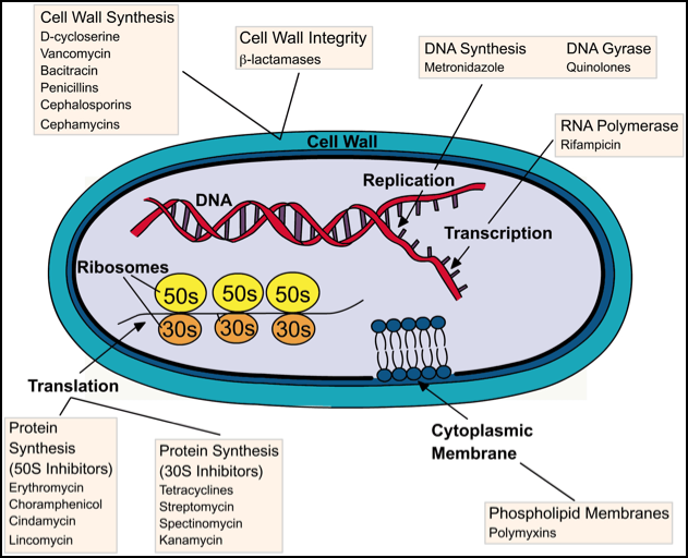
    Cell Wall Synthesis Inhibitors

    Sunday, February 7, 2016

    Pulmonology Review for Rotation Exams

    Pulmonology for Rotation Exams
    Peak Expiratory Flow - use a peak flow meter
    • If <350 L/min, perform PFTs to screen for obstruction
    Pulmonary Function Testing
    • FEV1: amount of air that can be forced out of the lungs in 1 second
      • Airway obstruction diagnosed by: normal/increased TLC with decreased FEV1
        • FEV1/FVC < 0.7
    Disease
    FEV1
    FVC
    FEV1/FVC
    FEF25-75
    FET
    Peak Exp Flow
    Obstructive
    Decreased
    <80%
    Normal
    <80%
    Decreased
    < 0.7
    Decreased
    <60%
    Increased
    Low
    Restrictive
    Normal or
    Decreased
    <80%
    Decreased
    <80%
    Normal or Increased
    0.7
    Normal
    >60%
    Normal
    Normal
    Mixed
    Decreased
    <80%
    Decreased
    <80%
    Decreased
    < 0.7
    Decreased or Normal
    <60%
    Increased or Normal

      • Tiffeneau index (FEV1/FVC x 100): % of FVC expired in one second
      • FET = Forced Expiratory Time
    • Pay Attention Here: The important thing to know about how to differentiate an obstructive vs. restrictive lung disease is based on their TLC, not the vital capacity, which will be decreased in obstructive lung diseases (this can be misleading).
    Disease
    TLC
    FRC
    TV
    RV
    VC
    Obstructive
    Increased
    Increased
    N
    Increased
    Decreased
    Restrictive
    Decreased
    Decreased
    N
    Decreased
    Decreased
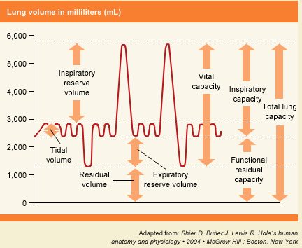
    • Total Lung Capacity: volume of air in lungs after maximum inspiration
    • Functional Residual Capacity: volume of air in lungs after normal expiration
    • Residual Volume: volume of air in the lungs at maximal expiration
    • Tidal Volume: volume of air breathed in and out of lungs during quiet breathing
    • Vital Capacity: volume of air expelled from the lungs during a maximum expiration
    1221_lung_volume.gif